Biophotonics and Biofluid Research Lab
Optical Coherence Tomography
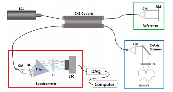
Optical coherence tomography (OCT) is an emerging optical imaging modality in biomedical optics and medicine. OCT performs high resolution, cross-sectional imaging of the internal microstructure and blood flow in biological tissues by measuring echoes of backscattered light as shown in Fig. 1. Tissue pathology can be imaged in situ and in real time with resolutions of 1–15 μm, one to two orders of magnitude finer than conventional ultrasound (see a comparison chart shown in Fig. 2). The unique features of OCT make it a powerful imaging modality, which promises to enable many fundamental research and clinical applications.

Fig. 2. Comparison to other medical imaging methods.
OCT performs cross-sectional imaging by measuring the magnitude and echo time delay of backscattered light. Cross-sectional images are generated by performing multiple axial measurements of echo time delay (longitudinal scans or A-scans) and scanning the incident optical beam transversely, as shown in Fig. 3. This produces a two-dimensional data set, which represents the optical backscattering in a cross-sectional plane through the tissue. Images, or B-scans, can be displayed in false color or grey scale to visualize tissue pathology. Three-dimensional, volumetric data sets can be generated by acquiring sequential cross-sectional images by scanning the incident optical beam in a raster pattern. Three-dimensional OCT (3D-OCT) data contain comprehensive volumetric structural information and can be manipulated similar to MR or CT images.

Fig.3 Scanning method to construct a 2D or 3D tomographic image
OCT is a powerful imaging technology in medicine because it performs “optical biopsy;” the real time, in situ visualization of tissue microstructure, without the need to remove and process specimens. OCT has applications in several general clinical situations: (1) Where standard excisional biopsy is hazardous or impossible. Applications include tissues such as the eye, arteries, or nervous tissues; (2) Where standard excisional biopsy has sampling error. Excisional biopsy and histopathology are the standard for diagnosis of many diseases including cancer; however, if the excisional biopsy misses the lesion, a false negative occurs. OCT can guide excisional biopsy to reduce the number of biopsies required and to improve sensitivity by reducing sampling errors; (3) For guidance of interventional procedures. The ability to see beneath the tissue surface enables the guidance of procedures such as stent placement or atherectomy, as well as microsurgical procedures such as vessel and nerve anastomoses. Coupled with catheter, endoscopic, laparoscopic, or needle delivery devices, OCT promises to have a powerful impact on many medical applications ranging from the diagnosis of neoplasia, to enabling new minimally invasive surgical procedures. The development of functional extensions of OCT enables imaging and measurement of properties such as Doppler flow, displacement, birefringence, and spectral properties. (From Introduction to Optical Coherence Tomography by J. Fujimoto and W. Drexler)